2. 中国海洋大学水产学院 青岛 266003;
3. 江苏中洋集团 南通 226600
2. Ocean University of China, Fisheries College, Qingdao 266003 ;
3. Jiangsu Zhongyang Group, Nantong 226600
在鱼类早期发育过程中,其组织、器官及系统的发生发育伴随着重要结构和功能的改变,包括各项生理机能的从无到有,从不完善到完善的变化(Sánchez-Amaya et al, 2007; Padrós et al, 2011; 宋炜等, 2012),这些结构功能的出现对鱼类早期适应外界恶劣的生活环境,提高其存活率至关重要。在大多数鱼类仔鱼阶段,眼睛作为主要的感觉器官,在摄食、熟悉环境、逃避敌害、集群和洄游等活动中发挥着不可代替的作用(赵传絪等, 1989; 柴毅等, 2007)。深入研究不同发育时期鱼类视觉器官的发生发育,对了解其生理活动、生态适应性、仔鱼开口时间、最佳投喂时间和饵料选择上具有重大意义(胡先成, 2007)1)。目前,国内外关于鱼类眼睛视网膜结构和视觉特性的研究已有较多报道(柴毅等, 2007; 马爱军等, 2007; 史玲玲等, 2013; 李超等, 2014; Torisawa et al. 2007; Ali et al, 1977; Yúfera et al, 2014),而对于鱼类早期眼睛各个组织的发生发育研究报道极少。
1) 胡先成.河川沙塘鳢(Odontobutis potamophila)早期发育过程中的器官发生、营养代谢及其能量收支的研究.华东师范大学博士研究生学位论文, 2007, 26-35
美洲鲥学名美洲西鲱(Alosa sapidissima),属鲱形目(Clupeiformes)、鲱科(Clupeidae)、西鲱属(Alosa),主要分布在北美洲大西洋西岸、太平洋海岸的加州圣地亚哥到阿拉斯加及亚洲东南部等地区(Jordan et al, 1898; Jones et al, 1978; Moyle, 1976),典型的溯河产卵鱼类。美洲鲥肉质鲜嫩,丰腴肥美,含有丰富的必需氨基酸、不饱和脂肪酸、矿物质和微量元素(顾若波等, 2007),口感和味道可以与我国濒临灭绝的鲥(Tenualosa reevesii)相媲美,具有较高的营养价值,深受消费者青睐。自2003年美洲鲥引入我国至今,科研工作者已开展了大量的繁育攻关工作,养殖技术日益成熟,取得了较大的成果。目前,国外对美洲鲥繁育(Marcy et al, 1972; Shardo, 1995)、遗传多样性(Hasselman et al, 2013)及捕捞迁徙(Hasselman et al, 2013; Bethoney et al, 2013)等方面进行了研究,而国内主要集中在早期发育(Quinn et al, 1996; 张呈祥等, 2010; 洪孝友, 20111))及苗种培育(彭景书等, 2012; 吴文化等, 2004; 潘庭双等, 2006)等方面,对于美洲鲥早期发育过程中组织学研究报道匮乏(洪孝友, 2011)1)。本文通过对美洲鲥早期不同发育阶段眼睛组织学研究,旨在阐明美洲鲥视觉早期发生发育的机理,探讨其与摄食的关系,丰富美洲鲥早期发育的基础生物学资料,为开展美洲鲥人工苗种繁育和养殖提供相应的理论知识。
1) 洪孝友.美洲鲥早期发育的形态学及组织学观察.上海海洋大学硕士研究生学位论文, 2011, 31-40
1 材料与方法实验所用美洲鲥来自江苏中洋集团繁育中心,实验于2014年5月1日在江苏中洋集团进行。选取体质健壮、性成熟的3-4龄个体于产卵池中(700 m3),取同一批次自然受精卵置于锥形流水孵化缸中孵化,水流速为15-25 L/min,微量充气,保证卵上下浮动。孵化水温为(21.0±0.50)℃,每隔一段时间吸出坏死长霉卵。显微镜(NIKON YS100)下连续观察发育特征,记录发育时间,并在各个发育时期分别取100粒胚胎样品。孵化后仔鱼转移到室内5 t水泥池(2.5 m×2 m× 1.0 m)中。仔鱼3日龄开口,3-15日龄投喂淡水轮虫[优势种群为萼花臂尾轮虫(Brachionus calyciflorus)],密度为12-15个/ml;12-20日龄,添加桡足类和枝角类无节幼体,密度为15-20个/ml;20-35日龄,开始投喂大型桡足类和枝角类;自30日龄起,开始投喂山东升索微粒子颗粒饲料。仔稚鱼培育水温为(20.5±1.0)℃,溶解氧控制在6.0-7.5 mg/L, 氨氮控制在0.05 mg/L以下。每日定时清污,日换等温淡水1/3,培育用水为曝气3 d以上地下深井水。仔鱼孵化后至15日龄每天连续取样,16-42日龄,每隔1 d取样1次,45日龄取样1次。每次随机取样60尾,其中,15尾用于全长和眼径的测量,其余45尾和上述所取受精卵样本分别置于Bouin’s固定液中,室温固定24 h,70%乙醇多次换洗后保存备用。
取各个发育时期胚胎30粒,鱼苗15尾,常规乙醇梯度脱水、二甲苯透明及石蜡包埋,运用LeiCa RM2265轮转式切片机进行连续切片,切片厚度为4-5 mm,HE染色,中性树胶封片,NIKON YS100显微镜下观察拍照,应用Photoshop 5.0软件优化处理图片。
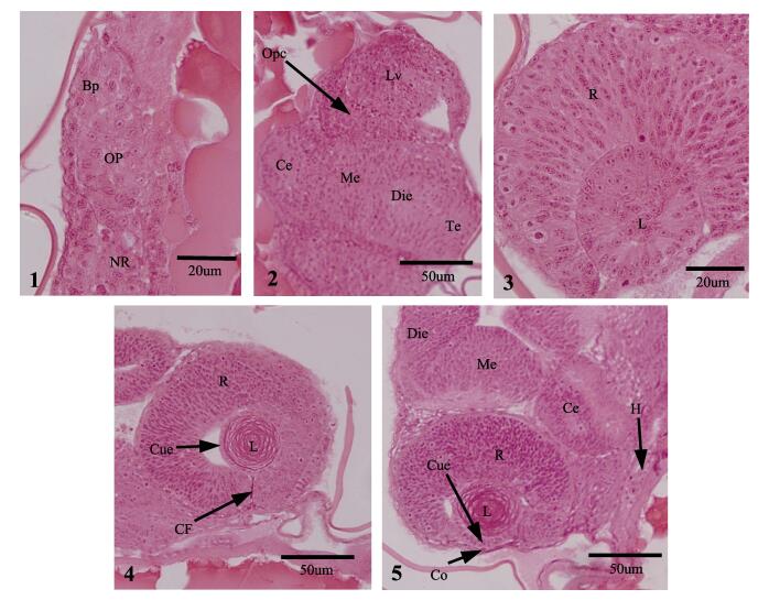
2 结果 2.1 美洲鲥胚胎期眼睛的发育受精21 h 13 min时,胚盘下包90%,神经褶在前端愈合膨大形成脑原基,在脑两侧区域形成对称的囊状突起即视泡(图 1-1)。

|
图 1 美洲鲥胚胎期眼睛的组织发育 Figure 1 The histological development of the eye in American shad at the embryonic stage 1.受精后21 h 13 min,视泡出现;2.受精后26 h 23 min,视杯形成,可见晶状体泡;3.受精后35 h 44 min,原始视网膜形成,晶状体开始分化;4.受精后47 h 35 min,可见晶状体立方上皮,脉络膜裂出现;5.受精后60 h 15 min,巩膜形成,可见晶状体纤维;Bp, 脑原基;Ce, 小脑;CF, 脉络膜裂;Co, 角膜;Cue, 立方上皮;Die, 间脑;H, 心脏;L, 晶状体;Lv, 晶状泡;Me, 中脑;NR, 神经索;OP, 视泡;Opc, 视杯;R, 视网膜;Te, 端脑 1. The optic vesicle was observed at 21 h 13 min post-fertilization; 2. The optic cup and lens vesicle were visable at 26 h 23 min post-fertilization; 3. The primitive retina appeared and the lens began to differentiate at 35 h 44 min post-fertilization; 4. The cubical epithelium of lens was visible and choroid fissure appeared at 47 h 35 min post-fertilization; 5. The cornea became formed and lens fibers were obvious at 60 h15 min post-fertilization; Bp, brain primordium; Ce, cerebellum; CF, choroid fissure; Co, cornea; Cue, cubical epithelium; Die, diencephalon; H, heart; L, lens; Lv, lens vesicle; Me, mesencephalon; NR, neural rod; OP, optic vesicle; Opc, optic cup; R, retina; Te, telencephalon |
受精26 h 23 min,胚体进入器官分化期,脑在形态上分化成了端脑、前脑、中脑和后脑,此时视泡外皮向内凹陷,形成双层杯状结构即视杯;贴近视杯的表皮外胚层逐渐增厚内陷入视杯形成晶状体泡(图 1-2)。
受精35 h 44 min,耳囊、心脏原基出现,尾芽开始偏离卵黄囊。视杯内外两层细胞不断靠拢生长、增厚,最终两层腔缝消失,外细胞层发育成为视网膜色素上皮,内细胞层发育成为原始的视网膜。此时,晶状体泡开始分化,晶状体内层细胞不断向内生长、延伸,在中心部位聚集,形成原始晶状体(图 1-3)。
受精47 h 35 min,心脏跳动频繁,胚胎肌肉抽动。晶状体被单层立方上皮所包裹,其内皮细胞增生、变长,继续向内延伸,形成初级晶状体纤维。在眼腹面可见明显的脉络膜裂(图 1-4)。
受精60 h15 min,仔鱼出膜前期,胚胎转动明显,鳍原基全部出现,卵黄囊斑点状色素明显。角膜在视网膜前缘的瞳孔处形成,由单一的立方上皮构成。晶状体新生纤维继续向中央环绕(图 1-5)。
2.2 美洲鲥胚后眼睛的发育美洲鲥受精卵在(21.0±0.5)℃条件下,经过70 h 15 min大量孵化出膜。初孵仔鱼全长为(7.45±0.15) mm,眼径为(0.38±0.03) mm,此时,仔鱼眼不具色素,视网膜最内层细胞最先分化出来,形成神经节细胞,细胞排列不规则,多呈长圆形。新生晶状体纤维逐层添加到原有纤维外侧,晶状体体积不断增大(图 2-1)。

|
图 2 美洲鲥胚后期眼睛的组织发育(Ⅰ) Figure 2 The histological development of the eye in American shad at post-embryonic stage (Ⅰ) 1.初孵仔鱼可见明显的晶状体、分化出的神经节细胞层;2. 1日龄仔鱼显示分化的视网膜;3. 1日龄仔鱼角膜分化为上皮和内皮细胞层;4. 2日龄仔鱼可见明显的素色层、纯视锥细胞的视觉层、脉络膜和由软骨组织和纤维层组成的巩膜;5. 2日龄仔鱼视网膜;6. 3日龄仔鱼视杆细胞和视网膜神经纤维出现;7. 3日龄仔鱼可见单层环状的虹膜;8. 5日龄仔鱼虹膜可见前缘层、基质层和色素层;9. 6日龄仔鱼脉络膜内可见血细胞;AC, 无长突细胞层;Bc, 血细胞;BC, 双极细胞层;Ca, 软骨组织;Cc, 视锥细胞;Cen, 角膜内皮;Cep, 角膜上皮;Ch, 脉络膜;Cue, 立方上皮;Fi, 纤维;G, 神经节细胞层;HC, 水平细胞层;IN, 内核层;IP, 内网膜层;Ip, 虹膜色素层;Ir, 虹膜;Ira, 虹膜前缘层;Irs, 虹膜基质层;L, 晶状体;NF, 神经纤维;Olm, 外界层;ON, 外核细胞层;OP, 外网膜层;P, 色素层;R, 视网膜;Rc, 视杆细胞;Sc, 巩膜;Vc, 视觉细胞层 1. Prominent lens and the ganglion cell layer was clearly identified in the developing retina of larvae at hatching; 2. The cornea differentiated into epithelial and endothelial cells on Day 1 after hatching (DAH); 3. Larvae on DAH 1 possessed differentiated retinal layers; 4. Larvae on DAH 2 showed an obvious pigmented retina with a visual cell layer including cones only, and the choroid appeared surrounding the retina and the sclera consisted of cartilage and outer fibrous layer; 5. Larvae on DAH 2 had clearly differentiated retina; 6. The rod cells were visible and the nerve fibrous layer was formed on DAH 3; 7. Larvae on DAH 3 showed the single annular iris; 8. The iris differentiated into three parts: iris anterior, stroma and pigment on DAH 5; 9. Blood cells in the choroid were visible on DAH 6. AC, amacrine cell layer; Bc, blood cells; BC, bipolar cell layer; Ca, cartilage; Cc, cone cells; Cen, corneal endothelium; Cep, corneal epithelium; Ch, choroid; Cue, cubical epithelium; Fi, fibre; G, ganglion cell layer; HC, horizontal cell layer; IN, inner nuclear layer; IP, inner plexiform layer; Ip, iris pigment; Ir, iris; Ira, iris anterior; Irs, iris stroma; L, lens; NF, nerve fibre; Olm, outer limiting membrane; ON, outer unclear layer; OP, outer plexiform layer; P, pigment; R, retina; Rc, rod cells; Sc, sclera; Vc, visual cell layer |
1日龄仔鱼全长为(7.91±0.23) mm,眼径为(0.39±0.01) mm,视网膜继续分化,在视网膜边缘出现一层薄薄的色素层,包含少量的黑色素颗粒;在视网膜色素层和神经节细胞层之间,可见排列紧密染色较深的外核层、厚度较宽细胞较大的内核层及着色较浅致密的内网膜层;聚集内部的晶状体纤维及其细胞核逐渐退化形成晶状体核,呈嗜酸性被HE染成深红色(图 2-2)。角膜立方上皮开始扁平化,视网膜外周的间充质细胞逐渐迁入预定角膜处和晶状体之间,在预定角膜处内侧形成一层薄的扁平细胞,即角膜内皮(图 2-3)。
2日龄仔鱼全长为(8.89±0.19) mm,眼径为(0.41±0.02) mm,巩膜形成,由软骨细胞层和弹力纤维组织构成,前端与角膜相连,与角膜共同包被整个眼球,起保护支撑作用;在巩膜和视网膜色素层之间,紧贴着巩膜内侧出现一层疏松的间充质组织,即脉络膜(图 2-4)。视网膜各层继续分化,最外色素层增厚明显,具大量的黑色素细胞。外界膜出现,由胶质细胞外侧突末端相互连接而成,分隔感光细胞内部与其细胞核。色素层与外核层之间出现视觉细胞层,为纯视锥细胞层,此时,色素层开始向视觉细胞层延伸。在外核层与内核层之间,形成染色较浅的薄层为外网膜层。内核层分化为3种神经细胞:最外侧的单层水平细胞、中部的双极细胞层和内部无长突细胞层(图 2-5)。
3日龄仔鱼全长为(9.20±0.15) mm,眼径为(0.42±0.03) mm,视网膜视觉细胞层出现视杆细胞,色素层扩展到视觉细胞层,遮蔽了部分视觉细胞的外段。视网膜神经纤维层形成,由外层的神经节细胞轴突组成,视神经纤维聚集成束形成视神经。在视神经纤维内侧内界膜形成,由胶质细胞内侧突末端相互连接而成,将视神经纤维和玻璃体分开,此时美洲鲥仔鱼视网膜分化完全,由自外向内的色素层、视觉细胞层、外界膜、外核层、外网膜层、内核层、内网膜层、视神经节细胞层、视神经纤维层和内界膜10层构成(图 2-6)。脉络膜开始向眼前缘延伸,在靠近角膜和瞳孔处形成1层薄薄的环形膜,即为虹膜,膜内含有零星的色素细胞(图 2-7)。
5日龄仔鱼全长为(9.52±0.29) mm,眼径为(0.45±0.01) mm,虹膜开始分化,虹膜基质层出现,由含有少量色素颗粒的疏松结缔组织构成;在预定虹膜处间充质细胞在基质层外侧发育为一层胶原纤维样结构,即前缘层;视网膜色素上皮向晶状体方向延伸,紧贴虹膜基质层形成虹膜色素层(图 2-8)。
6日龄仔鱼全长为(9.77±0.16) mm,眼径为(0.47±0.03) mm,脉络膜出现毛细血管,可见少量血细胞,为视网膜外侧区域提供营养;晶状体上皮不断增生、变长、形成新的次级晶状体纤维添加到晶状体核周围,晶状体纤维化明显(图 2-9)。
11日龄仔鱼全长为(11.83±0.32) mm,眼径为(0.51±0.04) mm,角膜分化明显,此时,角膜上皮发育为复层扁平上皮。在角膜内皮和复层扁平上皮之间分化出一层透明均质的薄膜,即前弹性膜,其内含微细的胶原原纤维和基质,不具再生能力。角膜后缘内皮细胞沿着虹膜向内延伸形成虹膜内皮层。晶状体可见明显的螺旋状纤维纹脉(图 3-1)。

|
图 3 美洲鲥胚后期眼睛的组织发育(Ⅱ)
Figure 3 The histological development of the eye in American shad at post-embryonic stage (Ⅱ)
1. 11日龄仔鱼所示角膜的复层扁平上皮、前弹性膜和内皮层,虹膜内皮层,晶状体纤维;2. 13日龄仔鱼脉络膜银膜出现;3. 14日龄仔鱼角膜完全分化;4. 16日龄仔鱼包含4层的虹膜;5. 16日龄仔鱼所示被HE染成黄色的晶状体核;6. 20日龄仔鱼所示明显的虹膜后缘层;7. 22日龄仔鱼所示脉络膜腺体和线状银膜;8. 26日龄稚鱼所示完全发育的虹膜;9. 45日龄幼鱼所示2层水平细胞;Ac.前房;Ar, 银膜;Ce, 柱状上皮细胞;Cen, 角膜内皮;Ch, 脉络膜;Chg, 脉络膜腺;Co, 角膜;Cos, 角膜基质层;CT, 疏松结缔组织;HC, 水平细胞层;Ip, 虹膜色素层;Ir, 虹膜;Ira, 虹膜前缘层;Ire, 虹膜内皮层;Irp, 虹膜后缘层;Irs, 虹膜基质层;L, 晶状体;Lea, 前弹性膜;Lep, 后弹性膜;LF, 晶状体纤维; MF, 肌肉纤维;ON, 外核细胞层;Pc, 后房;Ste, 复层扁平上皮;Vc, 视觉细胞层 1. Stratified squamous epithelium, lamina elastic anterior, endothelium in the cornea, the endothelium of the iris and the lens fibers were observed in larvae on DAH 11. 2. Argenteum appeared in the larvae on DAH 13; 3. Larvae on DAH 14 showed fully-developed cornea; 4. Larvae on DAH 16 possessed developing iris including four layers; 5. Larvae on DAH 16 showed yellow-colored lenticular nucleus stained with HE; 6. Larvae on DAH 20 showed obvious iris posterior; 7. Larvae on DAH 22 showed clear choroid gland and linear argenteum; 8. Fry on DAH 26 showed the fully-developed iris; 9. Juveniles on DAH 45 showed two layers of horizontal cells. Ac, anterior chamber; Ar, argenteum; Ce, columnar epithelium; Cen, corneal endothelium; Ch, choroid; Chg, choroid gland; Co, cornea; Cos, cornea stroma; CT, loose connective tissue; HC, horizontal cell layer; Ip, iris pigment; Ir, iris; Ira, iris anterior; Ire, iris endothelium; Irp, iris posterior; Irs, iris stroma; L, lens; Lea, lamina elastica anterior; Lep, lamina elastica posterior; LF, lens fibre; MF, muscle fibre; ON, outer unclear layer; Pc, posterior chamber; Ste, stratified squamous epithelium; Vc, visual cell layer |
13日龄仔鱼全长为(12.58±0.74) mm,眼径为(0.54±0.01) mm,脉络膜外层银膜出现(图 3-2)。
14日龄仔鱼全长为(12.85±1.28) mm,眼径为(0.62±0.05) mm,角膜的后弹性层由角膜内皮分泌形成,为一层透明的均质膜,较前弹性层薄,其具有弹性可再生。在前弹性层和后弹性层之间为角膜基质层,由粗细一致的胶原原纤维平行排列而成,内含角膜细胞。此时角膜分化完全,由自外向内的复层扁平上皮、前弹性层、基质层、后弹性层和内皮层5层构成(图 3-3)。
16日龄仔鱼全长为(15.00±1.76) mm,眼径为(0.66±0.02) mm,虹膜分化明显,基质层含有大量色素细胞,虹膜色素层上皮细胞呈现矮柱状;虹膜与角膜之间的腔隙称为前房,与晶状体之间的腔隙称为后房(图 3-4)。晶状体内部纤维层经HE染色变为黄色。巩膜外侧有丰富的肌肉层(图 3-5)。
20日龄仔鱼全长为(16.25±1.24) mm,眼径为(0.78±0.04) mm,虹膜继续分化,虹膜后缘层出现,为一层无细胞结构的透明膜(图 3-6)。
22日龄仔鱼全长为(18.05±1.74) mm,眼径为(0.92±0.08) mm,银膜继续增厚、延伸,呈现发达的线条形覆盖整个脉络膜,此时脉络膜结缔组织富含血管和色素颗粒;在眼背后部,银膜与血管膜之间,靠近视神经区域,可见明显的脉络膜腺(图 3-7)。
26日龄稚鱼全长为(21.3±1.55) mm,眼径为(1.15±0.05) mm,脉络膜色素贯穿整个脉络膜结缔组织层,此时脉络膜发育完全,由外向内依次为银膜层、血管层和色素层。虹膜基质层出现大量的肌肉纤维和色素,虹膜后缘层可见明显的色素颗粒,虹膜色素层继续向内延伸,富含大量的黑色素,其最外层为一层柱状细胞(图 3-8)。此时虹膜分化完全,由外向内依次为虹膜内皮层、前缘层、基质层、后缘层和色素层。45日龄幼鱼视网膜内核层具2层水平细胞(图 3-9)。此时,美洲鲥眼睛各个组织结构分化发育完全。
3 讨论在美洲鲥视觉器官发育过程中,视泡在原肠晚期既已出现,为对称的囊状突起。受精后35 h 44 min,原始视网膜和晶状体形成。这与大多数硬骨鱼类的发育时序相同(何滔等, 2012; 佟雪红等, 2011; 胡先成等, 2007)。由此可知,视网膜和晶状体的早期分化对鱼类早期视觉的形成至关重要。美洲鲥初孵仔鱼视网膜无色素和视觉细胞。2日龄仔鱼巩膜形成,由软骨组织和外围的纤维组成。3日龄仔鱼视网膜分化明显,具10层结构,原始脉络膜和虹膜出现,此时仔鱼已经具备了初始视觉成像系统。眼睛的早期分化和发育为其开口摄食提供了视觉上的保证。13日龄脉络膜外层银膜出现,其为鱼类所特有的,内含鸟粪素,可将射入眼球的微弱光线反射到视网膜上,这个结构的出现为仔鱼更好的适应弱光环境提供了保障。14日龄仔鱼角膜分化发育完全,此时仔鱼眼睛具备了较强的屈光能力,对外界刺激敏感。22日龄脉络膜腺出现,它对来自心脏的血液所产生的压力起缓冲作用,可以减少对视网膜的机械损伤,具有保护视网膜的作用。26日龄仔鱼脉络膜和虹膜分化发育完善,使外界反射的物象更加清楚,此时稚鱼具备完善的遮光、调节能力,活动更加敏捷。美洲鲥早期眼睛发育特点与条石鲷(Oplegnathus fasciatus)、河川沙塘鳢(Odontobutis potamophila)类似,但在发育时序上具有一定的差别(何滔等, 2012; 胡先成等, 2007)。
视网膜是形成视影像的重要组成部分,外界光能通过视网膜形成电信号,沿着视路传递到视觉中枢而形成视觉。视觉是鱼类进行摄食活动的重要感觉器官之一,其在摄食行为中主要体现在寻找和发现、辨认和选择、摄食方向和姿势调整(李大勇等, 1994)。美洲鲥2日龄仔鱼视网膜可见明显的视觉细胞层,由视锥细胞组成,这与其他鱼类的报道相同(柴毅等, 2007; 胡先成等, 2007; 史玲玲等, 2013; Wahl et al, 1993)。此时,美洲鲥仔鱼初具游泳能力,位于水层中上部,适于感应强光。3日龄稚鱼视网膜分化完全,具备10层结构,由外向内依次为色素层、视觉细胞层、外界膜、外核层、外网膜层、内核层、内网膜层、视神经节细胞层、视神经纤维层和内界膜,而软骨鱼施氏鲟视网膜仅分化为8层结构,缺少内、外界膜结构(史玲玲等, 2013),可见美洲鲥眼睛的结构分化更加复杂,在功能上可能强于施氏鲟。另外,高小强等(2015)研究发现,美洲西鲱仔鱼3日龄开口,初次摄食率高达46.6%,且美洲西鲱不同发育阶段一昼夜中摄食高峰存在差异,即4日龄仔鱼一昼夜具有1个摄食高峰,17日龄仔鱼一昼夜中具有两个摄食高峰,而28日龄稚鱼一昼夜则具有3个摄食高峰。结合本研究结果,美洲鲥仔鱼开口摄食时,视网膜已经分化完全,且视觉细胞层视杆细胞出现,使仔鱼具备了感受弱光的能力,从而为初次摄食提供了有效的保证;此后随着发育的进行,视杆细胞逐渐增多,角膜(14日龄)、虹膜(26日龄)和脉络膜(26日龄)的分化发育成熟,视觉系统逐渐发育完善,美洲鲥摄食能力不断得到加强,摄食成功率也不断提高。综上所述,美洲鲥的视觉发育与其摄食行为特性相适应。
不同生态习性的鱼类具备不同的视觉特性,如光感受系统发达的中上层鱼类具备4层水平细胞,而光感受系统不发达的底栖鱼类一般仅有1-2层水平细胞(Biaxter et al, 1970)。半滑舌鳎终生无分化的水平细胞(马爱军等, 2007),鳜仔鱼视网膜水平细胞仅为1层,属于光感受系统不发达种类(田文斐等, 2012)。潘庭双等(2006)研究表明,美洲鲥生殖洄游的亲鱼常活动于水体中下层,产卵后群体即行解散, 降河入海索饵和越冬,有些鱼苗在降河洄游时不会入海, 而只在海口深水层的洞穴里越冬。在本研究中,美洲鲥发育到幼鱼阶段内核层的水平细胞为两层,属于视觉系统不发达的鱼类,这与其生态习性相适应。
| 马爱军, 王新安, 庄志猛, 等. 半滑舌鳎仔、稚鱼视网膜结构与视觉特性. 动物学报 , 2007, 53 (2) : 354-363 | |
| 史玲玲, 危起伟, 柴毅, 等. 达氏鲟视网膜早期发育及其相关机能. 中国水产科学 , 2013, 20 (5) : 958-967 | |
| 田文斐, 钟俊生, 钱叶洲, 等. 鳜仔鱼视网膜及口腔齿的发育对摄食的适应. 上海海洋大学学报 , 2012, 21 (2) : 190-198 | |
| 李超, 王亮, 覃乐政, 等. 4种岩礁性鱼类视网膜感光细胞和最小分辨角的比较. 水产学报 , 2014, 38 (3) : 400-409 | |
| 李大勇, 何大仁, 刘晓春. 光照对真鲷仔、稚、幼鱼摄食的影响. 台湾海峡 , 1994, 13 (1) : 26-31 | |
| 宋炜, 宋佳坤. 西伯利亚鲟仔稚鱼胚后发育的形态学和组织学观察. 中国水产科学 , 2012, 19 (5) : 790-798 | |
| 张呈祥, 徐钢春, 徐跑, 等. 美洲鲥仔、稚、幼鱼的形态发育与生长特征. 中国水产科学 , 2010, 17 (6) : 1227-1233 | |
| 吴文化, 王斌, 师伟, 等. 美洲鲥鱼苗种规模化养殖技术研究Ⅰ.稚鱼培育. 水产学杂志 , 2004, 17 (2) : 61-64 | |
| 何滔, 肖志忠, 刘清华, 等. 条石鲷视觉器官早期发育的组织学观察. 海洋科学 , 2012, 36 (3) : 49-53 | |
| 佟雪红, 马道远, 徐世宏, 等. 大菱鲆(Scophthalmus maximus)胚胎发育的形态学和组织学研究. 海洋与湖沼 , 2011, 42 (6) : 844-849 | |
| 赵传絪, 唐小曼, 陈思行. 鱼类的行动. 北京: 农业出版社, 1989 : 263 . | |
| 胡先成, 赵云龙. 河川沙塘鳢视觉器官的发育及其与摄食的关系. 动物学杂志 , 2007, 42 (5) : 41-48 | |
| 顾若波, 张呈祥, 徐钢春, 等. 美洲鲥肌肉营养成分分析与评价. 水产学杂志 , 2007, 20 (2) : 40-46 | |
| 柴毅, 谢从新, 危起伟, 等. 中华鲟视网膜早期发育及趋光行为观察. 水生生物学报 , 2007, 31 (6) : 920-922 | |
| 高小强, 洪磊, 刘志峰, 等. 美洲西鲱仔鱼不可逆点及仔、稚鱼摄食特性研究. 水产学报 , 2015, 39 (3) : 392-400 | |
| 彭景书, 白成, 陶后全, 等. 美洲鲥仔稚鱼淡、海水培育试验. 广东海洋大学学报 , 2012, 32 (1) : 97-100 | |
| 潘庭双, 李海洋, 侯冠军. 美洲鲥鱼的生物学特性及大规格苗种培育技术. 水利渔业 , 2006, 26 (4) : 33-34 | |
| Ali MA, Anctil M. Retinal structure and function in the walleye (Stizostedion vitreum vitreum) and sauger (S. canadense). J Fish Res Board Can , 1977, 34 (10) : 1467-1474 DOI:10.1139/f77-211 | |
| Bethoney ND, Schondelmeier BP, Stokesbury KDE, et al. Developing a fine scale system to address river herring (Alosa pseudoharengus, A. aestivalis) and American shad (A. sapidissima) bycatch in the U.S. Northwest Atlantic mid-water trawl fishery. Fish Res , 2013, 141 (4) : 79-87 | |
| Biaxter JHS, Stalnes M. Pure-cone retinae and retinomotor responses in larval teleosts. J Mar Biol Ass UK , 1970, 50 (2) : 449-460 DOI:10.1017/S0025315400004641 | |
| Hasselman DJ, Ricard D, Bentzen P. Genetic diversity and differentiation in a wide ranging anadromous fish, American shad (Alosa sapidissima), is correlated with latitude. Mol Ecol , 2013, 22 (6) : 1558-1573 DOI:10.1111/mec.12197 | |
| Jones W, Martin FD, Hardy JD. Development of fishes of the mid-atlantic bight: an Atlas of egg, larval, and juvenile Stages. , 1978 : 98-104 | |
| Jordan DS, Evermann BW. . The fishes of north and middle America: A descriptive catalogue of the species of fish-like vertebrates found in the waters of North America, north of the Isthmus of Panama , 1898 : 427-428 | |
| Marcy BC. Spawning of the American shad, Alosa sapidissima, in the lower Connecticut River. Chesapeake Sci , 1972, 13 (2) : 116-119 DOI:10.2307/1351013 | |
| Moyle PB. . Inland fishes of California , 1976 : 405 | |
| Padrós F, Villalta M, Gisbert E, et al. Morphological and histological study of larval development of the Senegal sole Solea senegalensis: an integrative study. J Fish Biol , 2011, 79 (1) : 3-32 DOI:10.1111/j.1095-8649.2011.02942.x | |
| Quinn TP, Adams DJ, Dean JA. Environmental changes affection the migratory timing of American shad and sockeye salmon. Ecology , 1996, 77 (4) : 1151-1162 DOI:10.2307/2265584 | |
| Sánchez-Amaya MI, Ortiz-Delgado JB, García-López Á, et al. Larval ontogeny of redbanded seabream Pagrus auriga Valenciennes, 1843 with special reference to the digestive system. A histological and histochemical approach. Aquaculture , 2007, 263 (1-4) : 259-279 DOI:10.1016/j.aquaculture.2006.10.036 | |
| Shardo JD. Comparative embryology of teleostean fishes. Ⅰ. Development and Staging of the American Shad, Alosa sapidissima (Wilson, 1811). J Morphol , 1995, 225 (2) : 125-167 DOI:10.1002/(ISSN)1097-4687 | |
| Torisawa S, Takagi T, Ishibashi Y, et al. Changes in the retinal cone density distribution and the retinal resolution during growth of juvenile Pacific bluefin tuna Thunnus orientalis. Fish Sci , 2007, 73 (5) : 1202-1204 DOI:10.1111/fis.2007.73.issue-5 | |
| Wahl C, Mills E, McFarland WN, et al. Ontogenetic changes in prey selection and visual acuity of the yellow perch Perca flavescens. Can Fish Aqua Sci , 1993, 50 (4) : 743-749 DOI:10.1139/f93-085 | |
| Yúfera M, Ortiz-Delgado JB, Hoffman T, et al. Organogenesis of digestive system, visual system and other structures in Atlantic bluefin tuna (Thunnus thynnus) larvae reared with copepods in mesocosm system. Aquaculture , 2014, 426-427 : 126-137 DOI:10.1016/j.aquaculture.2014.01.031 |


