2. 水产科学国家级实验教学示范中心 上海海洋大学 上海 201306
2. National Demonstration Center for Experimental Fisheries Science Education, Shanghai Ocean University, Shanghai 201306
白斑综合征病毒(White spot syndrome virus,WSSV)是养殖对虾主要的病害之一,其传播范围广、致病力强,虾群感染3~7 d的死亡率高达90%~100%,给水产养殖业造成了巨大的经济损失(黄倢等, 1995; Lo et al, 1996; 马晓燕等, 2012; 何建国等, 1999)。研究表明,囊膜蛋白是介导病毒与宿主细胞发生相互作用的重要因子,在病毒侵染早期起关键作用,VP26、VP28和VP37是WSSV的主要囊膜蛋白,VP26存在于囊膜蛋白和核衣壳蛋白之间起联结作用(于力等, 2008);VP28与病毒抗原性有关,在病毒入侵宿主细胞过程中起重要作用(van Hulten et al, 2000、2001);VP37是粘附蛋白,可延缓WSSV的侵染过程(Liang et al, 2005; Liu et al, 2009)。
Ras基因是在生物进化过程中第一个被鉴定出来的非常保守的原癌基因,广泛存在于从酵母菌到人类的真核细胞中,因首先发现于大鼠肉瘤病毒(Rat sarcoma, Ras)而得名(Liu et al, 1995; Winston et al, 1996; Fan et al, 1997)。其分泌的RAS蛋白是偶联细胞表面受体与胞内效应因子的信号转导子,位于细胞膜内侧,属于小G蛋白家族,能结合GTP和GDP。结合GTP时为活性状态,能够与下游多种效应因子相互作用进行信号传递;当结合GDP时处于非活性状态,不能传递信号。所以,RAS蛋白在多种细胞跨膜受体介导的信号通路中起着分子开关的作用,在细胞增殖、分化、调亡、癌变等方面也起着非常重要的作用(Wennerberg et al, 2005; Winston et al, 1996; van der Weyden et al, 2007; Castro et al, 2005; Greenhough et al, 2007)。目前,关于对虾小G蛋白家族的研究多集中在Rab蛋白上,Praween等(2017)研究表明,在对虾感染传染性皮下及造血组织坏死病毒(IHHNV)和WSSV时,Rab5、Rab6、Rab7三种蛋白的表达量均上调,说明三者参与了对虾的先天性免疫防御系统。
本研究通过克隆表达获得凡纳滨对虾的RAS蛋白,利用Far-western技术在体外筛选出能与RAS蛋白相互作用的WSSV结构蛋白,并通过ELISA验证二者在数量上的相互作用关系,为深入全面地探索WSSV感染机制和防治措施奠定基础。
1 材料与方法 1.1 实验材料WSSV结构蛋白VP26、VP28N和VP37,表达载体pBAD/gⅢA由本实验室保存;TRIzol试剂、PrimeScript RT Reagent Kit with gDNA Eraser试剂盒、Ex Taq酶及DNA标准(DNA Marker DL2000)购自TaKaRa公司;E.coli Top10感受态细胞购自TIANGEN公司;氨苄青霉素(Amp+)、L-阿拉伯糖、质粒小提试剂盒购自Solarbio公司;T4 DNA连接酶、限制性内切酶NcoⅠ、XbaⅠ及蛋白Marker购自Thermo公司;地高辛(DIG)购自Roche公司;胶回收试剂盒购自ZYMO公司。
1.2 实验方法 1.2.1 凡纳滨对虾cDNA的合成剪取凡纳滨对虾的鳃丝,加入适量RNAiso充分研磨后,按照RNAiso试剂盒说明书提取RNA。依照PrimeScript RT Reagent Kit With gDNA Eraser试剂盒说明书将RNA反转录成cDNA。
1.2.2 凡纳滨对虾Ras编码区基因的克隆按照GenBank上的凡纳滨对虾Ras mRNA序列,设计特异性引物(Rass:5'-TACCATGGAGATGACGGAATA-CACG-3';Rasa:5'-ACTCTAGATCGAACACAATGC-ACTT-3',划线处为NcoⅠ和XbaⅠ酶切位点),以凡纳滨对虾鳃的cDNA为模板,PCR扩增Ras基因序列。PCR产物经1%的琼脂糖凝胶电泳后,回收纯化。
1.2.3 重组表达载体pBAD/gⅢA-Ras的构建PCR产物和pBAD/gⅢA载体经NcoⅠ和XbaⅠ双酶切,T4 DNA连接酶连接,构建重组表达载体,转化到E.coli Top10感受态细胞中,取100 μl转化液涂布到LB固体培养基(Amp+,50 μg/ml)上,37℃倒置培养。挑取单克隆,进行菌液PCR筛选阳性克隆,测序验证。
1.2.4 RAS生物学分析利用在线软件ProtParam tool分析蛋白的等电点和分子量,应用SignalP 3.0 Server预测氨基酸序列有无信号肽,利用NPS Network Protein Sequence Analysis在线软件,采用PHD方法预测RAS蛋白二级结构(王修芳等, 2016)。
1.2.5 RAS蛋白的表达测序正确的菌液用新鲜的LB培养液(Amp+,100 μg/ml)培养至OD600 nm为0.5~0.6,加入诱导剂L-阿拉伯糖(终浓度为0.2 g/L),37℃诱导4 h,对照组不加诱导剂。10000 r/min离心5 min,收集菌体,PBS缓冲液(pH=7.4)重悬,用超声破碎仪超声破碎后,10000 r/min离心30 min,分别收集诱导、未诱导菌液的上清液和沉淀,SDS-PAGE分析RAS蛋白表达情况,并将蛋白条带切下,送上海复旦大学进行蛋白质谱分析。
1.2.6 Co2+柱亲和层析纯化RAS蛋白诱导表达的菌液沉淀用A液(含6 mol/L盐酸胍,pH=7.4)溶解后,4℃ 10000 r/min离心10 min,收集上清液,依次经0.8 μm和0.45 μm滤膜过滤。向层析柱中加入树脂,PBS缓冲液(pH=7.4)平衡洗柱,A液将紫外吸收峰洗至0,加入蛋白上清液混匀,冰浴2 h,A液洗脱至峰值为0,C液(含150 mmol/L咪唑,pH=7.4)洗脱,并收集RAS蛋白。RAS蛋白经尿素梯度复性液(4、2、1、0 mol/L)透析复性后浓缩,SDS-PAGE检测蛋白纯化效果。
1.2.7 DIG标记RAS蛋白将DIG粉末溶于DMSO(1: 1000)中,与RAS蛋白充分混匀,4℃ PBS缓冲液(pH=7.4)过夜透析,以去除过量的DIG。
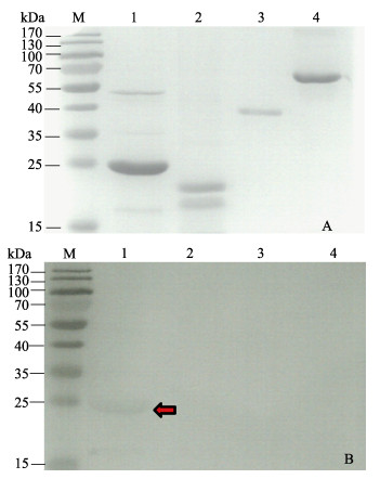
1.2.8 Far-western检测与RAS蛋白互作的WSSV蛋白将WSSV的3种结构蛋白VP26、VP28N、VP37和BSA(阴性对照)进行15% SDS-PAGE后,转印至PVDF膜上,4℃ 5% BSA封闭过夜,PBS-T清洗后加入DIG-RAS蛋白,室温避光孵育2 h,PBS-T清洗5遍后,加入碱性磷酸酶(AP)标记的抗DIG抗体(Anti-DIG-AP,1: 3000),室温避光孵育2 h,PBS-T清洗5遍后,用BCIP/NBT显色液避光显色。
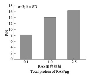
1.2.9 ELISA检测RAS蛋白与VP26的相互作用VP26用Na2CO3包被液稀释至0.02 μg/μl,分别取50 μl稀释好的VP26加入96孔板中,BSA为阴性对照,4℃包被过夜。PBS-T清洗后,5% BSA常温封闭2 h,PBS-T洗板3次。加入50 μl不同量的DIG-RAS蛋白(0.1、1.0、2.5 μg),常温孵育2 h,PBS-T洗板5次。加入50 μl Anti-DIG-AP(1: 5000稀释),常温孵育2 h,PBS-T洗板5次,加入50 μl PNPP显色液,直到出现明显颜色时,加入50 μl NaOH (0.3 mol/L)中止反应。酶标仪测定405 nm处的吸光值并计算P/N值。P/N计算公式:P/N=(OD405 nm-OD405 nm空白)/(OD405 nm BSA-OD405 nm空白)。
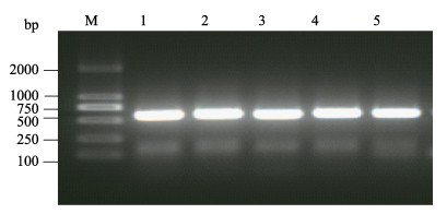
2 实验结果 2.1 Ras基因的克隆以凡纳滨对虾鳃的cDNA为模板,利用设计的引物扩增Ras基因,琼脂糖凝胶电泳结果见图 1,扩增的片段在500~750 bp之间,与Ras基因片段(561 bp)大小相符。
|
图 1 Ras基因的克隆 Fig.1 Amplification of Ras gene M:DNA标准品DL2000;1~5:Ras基因 M: DNA Marker DL2000; 1~5: Ras gene |
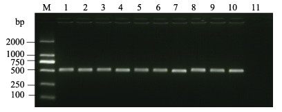
T4 DNA连接酶将双酶切后的目的片段与具有相同粘性末端的pBAD/gⅢA表达载体连接,转化到E. coli Top10细胞中,经菌液PCR鉴定阳性克隆,PCR产物电泳结果见图 2,条带大小为500~750 bp,菌液测序结果经过DNAMAN软件分析,重组表达载体序列完全正确,无碱基错配和移码突变。
|
图 2 重组表达载体PCR鉴定 Fig.2 Identification of recombinant vector by PCR M:DNA标准品DL2000;1~10: pBAD/gⅢA-Ras重组表达载体;11:阴性对照 M: DNA Marker DL2000; 1~10: Recombinant vector of pBAD/gⅢA-Ras; 11: Negative control |
经ProtParam tool分析显示,RAS理论分子量为21.3 kDa,等电点为5.97;SignalP 3.0 Server分析显示,该蛋白无信号肽结构,NPS Network Protein Sequence Analysis预测RAS蛋白二级结构(图 3),α螺旋为40.64%,β转角为36.9%,延伸链占22.46%。

|
图 3 RAS蛋白二级结构 Fig.3 Secondary structure prediction of RAS |
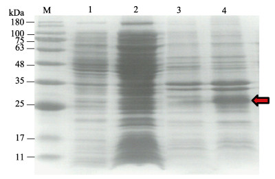
分别取诱导、未诱导菌液的上清液和沉淀进行SDS-PAGE(图 4),与未诱导的上清液、诱导的上清液、未诱导的沉淀相比,诱导菌液的沉淀在25 kDa左右出现一条明显加粗的蛋白条带(图 4中的红色箭头所示),将此条带进行蛋白质谱分析(表 1),结果显示,该蛋白为凡纳滨对虾的RAS蛋白。
|
图 4 RAS蛋白表达检测 Fig.4 Detection of RAS protein M:蛋白分子质量标准;1:未诱导上清液;2:诱导上清液;3:未诱导沉淀;4:诱导沉淀 M: Protein marker; 1: Supernatant protein of non-induced RAS; 2: Supernatant protein of induced RAS; 3: Pellet protein of non-induced RAS; 4: Pellet protein of induced RAS |
|
|
表 1 RAS蛋白质谱分析结果 Tab.1 Results of MS analysis of RAS |
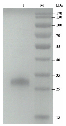
重组RAS蛋白带有6×His标签,可利用Co2+亲和层析法纯化。SDS-PAGE显示(图 5),RAS蛋白纯度高,为单一条带,可用于下一步实验。
|
图 5 SDS-PAGE分析纯化的RAS蛋白 Fig.5 SDS-PAGE analysis of purified RAS M:蛋白分子质量标准;1:纯化的RAS蛋白 M: Protein marker; 1: Purified RAS protein |
将VP26、VP28N和VP37进行蛋白电泳后转印至PVDF膜上,依次经过DIG-RAS蛋白和Anti-DIG-AP孵育。显色结果显示,在VP26泳道出现明显的条带(图 6中的红色箭头所示),而VP28N、VP37和BSA泳道无条带,说明RAS蛋白只能与VP26特异性结合。
|
图 6 Far-western分析RAS蛋白与VP26、VP28N及VP37的相互作用 Fig.6 Analysis of interaction of purified recombinand RAS and VP26, VP28N, and VP37 by far-western A:聚丙烯酰胺凝胶电泳;B:Far-western;M:蛋白分子质量标准;1:VP26;2:VP28N;3:VP37;4:BSA A: SDS-PAGE; B: Far-western; M: Protein marker; 1: VP26; 2: VP28N; 3: VP37; 4: BSA |
采用ELISA方法,进一步验证VP26与RAS互作,数据分析显示P/N值远大于2(图 7),且VP26与RAS蛋白的相互作用随RAS蛋白量的增加而增强,当RAS蛋白量增加至2.5 μg时,P/N值增加到16.4,说明VP26与RAS蛋白有明显的结合作用。
|
图 7 ELISA分析RAS蛋白与VP26的相互作用 Fig.7 Analysis of RAS interaction with VP26 by ELISA |
大量研究表明,病原蛋白与宿主蛋白相互作用是诱发感染、致病的关键环节。WSSV侵染宿主细胞是一个很复杂的过程,病毒在复制过程中会与多种宿主细胞蛋白发生作用。近年来,已报道多种宿主分子(包括Rab5、Rab6、Rab7等小G蛋白)参与WSSV的侵染过程(Sritunyalucksana et al, 2006、2013),但关于RAS蛋白与WSSV的相互作用尚不明确。
本研究根据Ras编码区序列设计引物,PCR获得Ras序列,通过原核表达和Co2+亲和层析法得到RAS蛋白,SDS-PAGE结果显示,RAS蛋白存在于诱导沉淀中,属于包涵体蛋白。以WSSV的3个主要结构蛋白VP26、VP28N和VP37作为研究对象,通过Far-western和ELISA检测三者是否与凡纳滨对虾RAS蛋白相互作用。研究表明,RAS蛋白虽不能与VP28N和VP37相互作用,但能与VP26特异性结合,且二者的相互作用随着RAS蛋白量的增加而增强。VP26是WSSV中含量较高的被膜蛋白,起连接作用(Tsai et al, 2006),既可以与囊膜蛋白VP28结合,又可以与核衣壳蛋白VP51结合,三者可能通过形成复合体起到稳定病毒结构的作用(Chang et al, 2008; Wan et al, 2008)。VP26能特异性结合斑节对虾(Penaeus monodon)血细胞,参与WSSV侵染的初始阶段(刘非等, 2006),中国明对虾(Fenneropenaeus chinensis)的Tetraspanin-3蛋白在体外也能特异地结合VP26,参与WSSV的感染过程(关广阔等, 2015),VP26通过与日本囊对虾(Penaeus japonicus)β肌动蛋白作用,完成WSSV的入侵、运输和释放等过程(Liu et al, 2011; Xie et al, 2005; Gouin et al, 2005),说明VP26可与多个宿主分子相互作用。
RAS是细胞内信号通路中的转导因子,在信号传导途径中起着极为重要的作用,RAS与VP28和VP37无作用,说明RAS不参与WSSV吸附宿主细胞的过程。根据研究结果可初步推测,在WSSV侵染凡纳滨对虾细胞时,RAS蛋白通过与VP26相互作用,调控信号通路介导WSSV进入细胞内,从而加快了WSSV在宿主体内的传播,具体作用机制尚需深入研究。本研究结果为进一步揭示WSSV侵染宿主细胞的信号通路和病毒-宿主相互作用机制提供了理论基础。
Castro AF, Rebhun JF, Quilliam LA. Measuring Ras-family GTP levels in vivo-running hot and cold. Methods, 2005, 37(2): 190-196 DOI:10.1016/j.ymeth.2005.05.015 |
Chang YS, Liu WJ, Chou TL, et al. Characterization of white spot syndrome virus envelope protein VP51A and its interaction with viral tegument protein VP26. Journal of Virology, 2008, 82(24): 12555-12564 DOI:10.1128/JVI.01238-08 |
Fan J, Bertino JR. K-ras modulates the cell cycle via both positive and negative regulatory pathways. Oncogene, 1997, 14: 2595-2607 DOI:10.1038/sj.onc.1201105 |
Gouin E, Welch MD, Cossart P. Actin-based motility of intracellular pathogens. Current Opinion in Microbiology, 2005, 8(1): 35-45 DOI:10.1016/j.mib.2004.12.013 |
Greenhough A, Patsos HA, Williams AC, et al. The cannabinoid δ9-tetrahydrocannabinol inhibits RAS-MAPK and PI3K-AKT survival signalling and induces BAD-mediated apoptosis in colorectal cancer cells. International Journal of Cancer, 2007, 121(10): 2172-2180 DOI:10.1002/(ISSN)1097-0215 |
Guan GK, Liu QH, Huang J. Interaction of tetraspanin-3 in Fenneropenaeus chinensis with WSSV in vitro. Progress in Fishery Sciences, 2015, 36(6): 56-62 [ 关广阔, 刘庆慧, 黄倢. 中国明对虾(Fenneropenaeus chinensis) Tetraspanin-3与WSSV的体外相互作用. 渔业科学进展, 2015, 36(6): 56-62] |
He JG, Zhou HM, Yao B, et al. White spot syndrome baculovirus (WSBV) host range and transmission route. Acta Scientiarum Naturalium Universitatis Sunyatseni, 1999, 38(2): 65-69 [ 何建国, 周化民, 姚伯, 等. 白斑综合症杆状病毒的感染途径和宿主种类. 中山大学学报(自然科学版), 1999, 38(2): 65-69 DOI:10.3321/j.issn:0529-6579.1999.02.015] |
Huang J, Song XL, Yu J, et al. Baculoviral hypodermal and hematopoietic necrosis—study on the pathogen and pathology of the explosive epidemic disease of shrimp. Marine Fisheries Research, 1995, 16(1): 1-10 [ 黄倢, 宋晓玲, 于佳, 等. 杆状病毒性的皮下及造血组织坏死——对虾暴发性流行病的病原和病理学. 海洋水产研究, 1995, 16(1): 1-10] |
Liang Y, Huang J, Song XL, et al. Four viral protein of white spot syndrome virus (WSSV) that attach to shrimp cell membranes. Diseases of Aquatic Organisms, 2005, 66(1): 81-85 |
Liu B, Tang X, Zhan W. Interaction between white spot syndrome virus VP26 and hemocyte membrane of shrimp, Fenneropenaeus chinensis. Aquaculture, 2011, 314(1-4): 13-17 DOI:10.1016/j.aquaculture.2011.01.023 |
Liu F, Cun SJ, Yang K, et al. Functional analysis on structure proteins VP28 and VP26 of white spot syndrome virus. Acta Scientiarum Naturalium Universitatis Sunyatseni, 2006, 45(4): 83-86 [ 刘非, 寸树健, 杨凯, 等. 对虾白斑综合症病毒(WSSV)结构蛋白VP28与VP26的功能分析. 中山大学学报(自然科学版), 2006, 45(4): 83-86 DOI:10.3321/j.issn:0529-6579.2006.04.020] |
Liu JJ, Chao JR, Jiang MC, et al. Ras transformation results in an elevated level of cyclin D1 and acceleration of G1 progression in NIH 3T3 cells. Molecular and Cellular Biology, 1995, 15(7): 3654-3663 DOI:10.1128/MCB.15.7.3654 |
Liu QH, Zhang XL, Ma CY, et al. VP37 of white spot syndrome virus interact with shrimp cells. Letters in Applied Microbiology, 2009, 48(1): 44-50 DOI:10.1111/lam.2009.48.issue-1 |
Lo CF, Ho CH, Peng SE, et al. White spot syndrome baculovirus (WSBV) detected in cultured and captured shrimp, crabs and other arthropods. Diseases of Aquatic Organisms, 1996, 27(3): 215-225 |
van der Weyden L, Adams DJ. The Ras-association domain family (RASSF) members and their role in human tumourigenesis. Biochimica et Biophysica Acta, 2007, 1776(1): 58-85 |
Ma XY, Li P, Yan H, et al. A review on shrimp white spot syndrome virus. Journal of Nanjing Normal University(Natural Science Edition), 2012, 35(4): 90-100 [ 马晓燕, 李鹏, 严洁, 等. 对虾白斑综合症病毒的概述. 南京师大学报(自然科学版), 2012, 35(4): 90-100 DOI:10.3969/j.issn.1001-4616.2012.04.017] |
Choubey PK, Roy JK. Rab11, a vesicular trafficking protein, affects endoreplication through Ras-mediated pathway in Drosophila melanogaster. Cell and Tissue Research, 2017, 367(2): 269-282 DOI:10.1007/s00441-016-2500-0 |
Sritunyalucksana K, Utairungsee T, Sirikharin R, et al. Virus- binding proteins and their roles in shrimp innate immunity. Fish & Shellfish Immunology, 2013, 33: 1269-1275 |
Sritunyalucksana K, Wannapapho W, Lo CF, et al. PmRab7 is a VP28-binding protein involved in white spot syndrome virus infection in shrimp. Journal of Virology, 2006, 80(21): 10734-10742 DOI:10.1128/JVI.00349-06 |
Tsai JM, Wang HC, Leu JH, et al. Identification of the nucleocapsid, tegument, and envelope proteins of the shrimp white spot syndrome virus virion. Journal of Virology, 2006, 80(6): 3021-3029 DOI:10.1128/JVI.80.6.3021-3029.2006 |
van Hulten MCW, Westenberg M, Goodall SD, et al. Identification of two major virion protein genes of white spot syndrome virus of shrimp. Virology, 2000, 266(2): 227-236 |
van Hulten MCW, Witteveldt J, Snippe M, et al. White spot syndrome virus envelop protein VP28 is involved in the systemic infection of shrimp. Virology, 2001, 285(2): 228-233 DOI:10.1006/viro.2001.0928 |
Wang XF, Liu QH, Wu Y, et al. cDNA clonging of coat-epsilon gene and its tissue distribution in Fenneropenaeus chinensis. Progress in Fishery Sciences, 2016, 37(4): 147-152 [ 王修芳, 刘庆慧, 吴垠, 等. 中国明对虾(Fenneropenaeus chinensis) coat-ε基因全长cDNA克隆及组织分布. 渔业科学进展, 2016, 37(4): 147-152] |
Wan Q, Xu L, Yang F. VP26 of white spot syndrome virus functions as a linker protein between the envelope and nucleocapsid of virions by binding with VP51. Journal of Virology, 2008, 82(24): 12598-12601 DOI:10.1128/JVI.01732-08 |
Wennerberg K, Rossman KL, Der CJ. The Ras superfamily at a glance. Journal of Cell Science, 2005, 118(5): 843-846 DOI:10.1242/jcs.01660 |
Winston JT, Coats SR, Wang YZ, et al. Regulation of the cell cycle machinery by oncogenic ras. Oncogene, 1996, 12(1): 127-134 |
Xie X, Yang F. Interaction of white spot syndrome virus VP26 protein with actin. Virology, 2005, 336(1): 93-99 |
Yu L, Li QZ. Advances of white spot syndrome virus. Chinese Journal of Preventive Veterinary Medicine, 2008, 30(6): 486-490 [ 于力, 李庆章. 虾白斑综合征病毒的研究进展. 中国预防兽医学报, 2008, 30(6): 486-490] |


