2. 上海海洋大学 水产种质资源发掘与利用教育部重点实验室 水产科学国家级实验教学示范中心 海洋动物系统分类与进化上海高校重点实验室 上海 201306;
3. 茂名市金阳热带海珍养殖有限公司 茂名 525444
2. Shanghai Ocean University, Key Laboratory of Exploration and Utilization of Aquatic Genetic Resources, Ministry of Education; National Demonstration Center for Experimental Fisheries, Science Education; Key Laboratory of Marine Animal Taxonomy and Evolution, Shanghai 201306;
3. Maoming Jinyang Tropical Fish Breeding Co. Ltd, Maoming 525444
鱼类的淋巴器官主要包括胸腺、头肾、脾脏以及黏膜相关的淋巴组织(Boehm et al, 2012),黏膜相关淋巴组织包括鼻咽、内脏、鳃和皮肤相关淋巴组织,它们通过细胞和体液介导途径参与继发性淋巴免疫反应。胸腺是大多数鱼类最早发育的中枢淋巴器官,它在鱼类胚胎发育早期就已经形成并发挥作用,胸腺为T细胞的增殖、成熟和抗原受体库的生成提供了适宜的微环境(Nakanishi et al, 2015),同时,产生促淋巴细胞生成素以促进其他淋巴器官的淋巴细胞生成(Fishelson, 2006)。头肾和脾脏是主要的免疫及造血器官,含有丰富的血管、血窦和免疫细胞(胡玲玲等, 2010)。其免疫细胞介导并参与免疫反应,尤其吞噬细胞(黑色素–巨噬细胞和粒细胞)是免疫系统的重要组成部分,具有趋化、吞噬、细胞因子分泌、抗原处理以及通过多种机制表达来清除病原体的作用(Espinosa et al, 2018; 邢贺飞等, 2016)。免疫淋巴器官是鱼类防止病原入侵的主要防线,研究免疫淋巴器官的发生发育可了解机体免疫活性建立的时间节点,对优化养殖条件、完善苗种培育和健康养殖技术具有重要意义(Campoverde et al, 2019)。近年来,国内外越来越多的学者针对鱼类免疫淋巴器官的发生发育开展研究,如在大西洋白姑鱼(Argyrosomus regius)、考氏鳍竺鲷(Pterapogon kauderni)、斑马鱼(Barchydanio rerio var)、牙鲆(Paralichthys olivaceus)、草鱼(Ctenopharyngodon idellus)和斜带石斑鱼(Epinephelus coioides)等多种鱼类已见报道(Campoverde et al, 2019; Fishelson, 2010; Danilova et al, 2004; Liu et al, 2004; 雷雪彬等, 2013; 吴金英等, 2003)。
四指马鲅(Eleutheronema tetradactylum)俗称马友、午鱼、午笋等,通常分布于热带和温带海区,主要分布于印度洋和太平洋西部,在我国黄海、东海、南海和渤海均有分布(Moore et al, 2011; Wang et al, 2014)。其肉质鲜美,营养价值高,具有良好的养殖前景。2012年我国规模化全人工繁育和养殖四指马鲅属鱼类获得成功,近年来先后在广东、海南、福建、广西、上海、江苏、山东、浙江、河北等沿海地区开展了养殖技术推广应用,四指马鲅已成为海水鱼类养殖的新品种(林先智等, 2015)。目前,四指马鲅苗种培育和养殖技术已经成功,但在仔稚鱼变态发育阶段常出现大量死亡现象,同时,在成鱼养殖过程中对应激反应比较敏感,容易因手工操作受伤死亡,其中原因需要进一步研究(刘奇奇等, 2017)。鱼类淋巴器官对病害具有重要的免疫作用,关于四指马鲅淋巴器官发生发育的研究目前在国内外尚未见报道。本研究采用石蜡组织连续切片和HE染色技术对人工培育的四指马鲅淋巴器官发育进行研究,试从免疫器官发育的角度推测机体免疫活性建立的时间,并观察淋巴器官发育过程中组织形态学特点,以丰富四指马鲅的基础生物学资料,为其苗种培育和成鱼健康养殖提供理论参考。
1 材料与方法 1.1 实验材料实验所用的四指马鲅仔稚鱼为2018年6~9月在中国水产科学研究院南海水产研究所珠海试验基地培育所得。
1.2 实验方法鱼苗孵化及培育参考区又君等(2017)的方法进行。孵化后2 dph (Days post hatching, dph)将鱼苗置于1000 L白色塑料桶中培育,放苗前,水桶中预先接种小球藻和轮虫,每天添加淡水降低盐度0.5~1,直到盐度降至9.0±0.5。培育水温为(28±2)℃。7 dph投喂枝角类和桡足类幼体,25 dph起投喂卤虫无节幼体,30 dph起投喂粉状配合饲料并逐渐过渡到人工颗粒饲料。1~ 18 dph,每天取样1次;20~45 dph,每5 d取样1次;45~ 60 dph,每7 d取样1次,每次取15尾。取样时,记录鱼苗日龄,并用数显游标卡尺(精确度0.01 mm)测量全长,置于4%多聚甲醛中固定,24 h后转入70%的乙醇中保存。参考马定昌等(2014)的方法并稍作调整,将固定好的样品进行流水冲洗12 h,经50%~100%梯度乙醇脱水(50%乙醇2 h、70%乙醇4 h、80%乙醇2 h、85%乙醇2 h、95%乙醇45 min×2、100%乙醇45 min×2),二甲苯透明(1/2无水乙醇+1/2二甲苯混合液1 h、二甲苯15 min×2),浸蜡(石蜡2 h×2),石蜡包埋,以4~6 μm的厚度进行纵向连续切片,苏木精‒伊红(HE)染色,中性树脂封片,在ZEISS Axio Scope. A1型光学显微镜(德国)下观察并摄影记录。
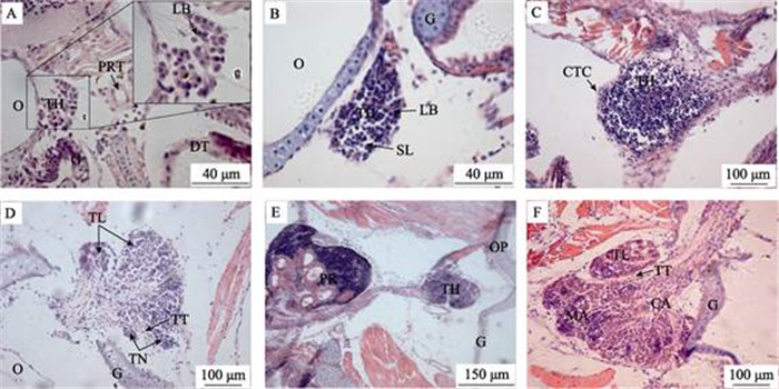
2 结果 2.1 胸腺3 dph,胸腺原基开始形成,位于鳃盖骨背上角处,由4~6层未分化干细胞和淋巴母细胞样的细胞组成,细胞较大,圆形或椭圆形,嗜碱性,核大且深染,原基外缘上皮鞘尚未形成(图 1A)。6 dph,胸腺个体增大,呈长椭圆形,胸腺细胞增多,染色加深,出现小淋巴细胞,呈紫红色,上皮鞘形成(图 1B)。18 dph,胸腺外包结缔组织被膜,淋巴细胞增多,密度增大(图 1C)。20 dph,结缔组织开始向胸腺实质伸展把腺体分成若干小叶,小叶间结缔组织形成的小梁将其分成数个小结(图 1D)。25 dph,胸腺与头肾有结缔组织连接(图 1E),胸腺分区明显,皮质区位于胸腺外层,淋巴细胞密集,着色较深,有许多微血管分布;髓质区位于胸腺内层,着色较浅,淋巴细胞数量较少。胸腺小叶明显,由于小叶间隔不明显,小结不完全隔开(图 1F)。胸腺主要由着色较深的淋巴母细胞和淋巴细胞所填充,偶见少量红细胞。

|
图 1 四指马鲅胸腺的发育
Fig.1 Development of thymus in E.tetradactylum
A:3dph (Days post hatching),示胸腺淋巴母细胞,400×;B:6 dph,示胸腺淋巴母细胞和小淋巴细胞,400×; C:18 dph,示胸腺淋巴细胞和结缔组织被膜,200×;D:20 dph,示胸腺小叶、小梁及小结出现,200×;E:25 dph, 示头肾和胸腺,100×;F:25 dph,示胸腺皮质区、髓质区、小叶、小梁,400×;CTC:结缔组织被膜;CA:皮质区; DT:消化道;G:鳃弓;LB:淋巴母细胞;MA:髓质区;O:耳囊;OP:鳃盖骨;PR:头肾;PRT:前肾管; SL:小淋巴细胞;TH:胸腺;TL:胸腺小叶;TT:胸腺小梁;TN:胸腺小结 A: 3 dph, showing lymphoblasts of the thymus, 400×; B: 6 dph, showing lymphoblasts and small lymphocytic of the thymus, 400×; C: 18 dph, showing lymphocytes and connective tissue, 200×; D: 20 dph, showing thymic lobules, trabeculae, and nodules appear, 200×; E: 25 dph, showing thymus and pronephros; F: 25 dph, showing the cortical areas, medulla areas, lobule, trabecular, 400×; CTC: Connective tissue; CA: Cortical areas; DT: Digestive tract; G: Gill; LB: Lymphoblasts; MA: Medulla areas; O: Oticcapsule; OP: Operculum; PR: Pronephros; PRT: Pronephric tubules; SL: Small lymphocytic; TH: Thymus; TL: Thymic lobule; TT: Thymic trabecular; TN: Thymic nodules |
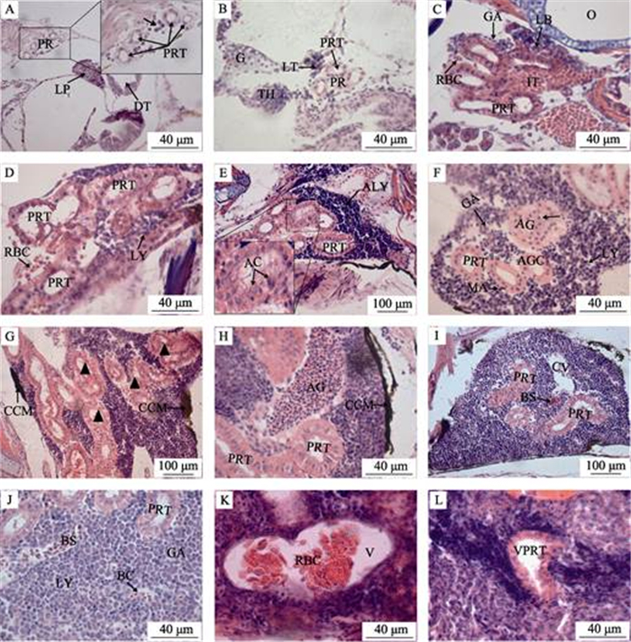
另外,在早期发育过程中,胸腺外周有一些分散的淋巴细胞,胸腺与头肾有细胞“桥”连接(图 2B), 到发育基本完成,细胞“桥”衍变成结缔组织,胸腺与头肾独立分开。

|
图 2 四指马鲅头肾的发育
Fig.2 Development of head kidney in E.tetradactylum
A:3 dph,示头肾的造血干细胞(↑)和前肾管,400×;B:5 dph,示增大的前肾管和淋巴组织,400×;C:7 dph,示继续增大的前肾管,管间组织,淋巴母细胞,粒细胞,红细胞,400×;D:15 dph,示前肾管,淋巴细胞,红细胞,400×;E:18 dph,示淋巴细胞集中区和退化的肾管,凋亡细胞,200×;F:20 dph,示肾上腺细胞,前肾管,粒细胞,淋巴细胞,巨噬细胞,400×;G:25 dph,示大量前肾管退化(▲),嗜铬细胞团,200×;H:30 dph,示前肾管,肾上腺,嗜铬细胞团,200×;I:35 dph,示中央静脉,血窦,前肾管,200×;J:40 dph,示前肾管,血窦,血细胞,淋巴细胞,粒细胞,400×;K:53 dph,示静脉,400×;L:53 dph,示已退化的前肾管,400×;AC:凋亡细胞;AG:肾上腺;AGC:肾上腺细胞;ALY:淋巴细胞集中区; BC:血细胞;BS:血窦;CCM:嗜铬细胞团;CV:中央静脉;DT:消化道;G:鳃弓;GA:粒细胞;IT:管间组织; LP:肝原基;LT:淋巴组织;LB:淋巴母细胞;LY:淋巴细胞;MA:巨噬细胞;ND:肾管; PR:前肾;PRT:前肾管;RBC:红细胞;V:静脉;VPRT:退化的前肾管 A: 3 dph, showing hematopoietic stem cells of head kidney(↑) and pronephric tubules, 400×; B: 5 dph, showing enlarged pronephric tubules and lymphatic tissue, 400×; C: 7 dph, showing pronephric tubules continues to enlarge, inter tubular tissue, lymphocytoblast, granulocytes, red blood cell, 400×; D: 15 dph, showing pronephric tubules, lymphocyte, red blood cell, 400×; E: 18 dph, showing lymphocytes accumulation, degenerated pronephric tubules and apoptotic cell, 200×; F: 20 dph, showing adrenal gland cells, pronephric tubules, granulocytes, lymphocyte, macrophage, 400×; G: 25 dph, showing a lot of pronephric tubules degenerate(▲), chromaffin cell mass, 200×; H: 30 dph, showing pronephric tubules, adrenal gland, chromaffin cell mass, 200×; I: 35 dph, showing central veins, blood sinus, pronephric tubules, 200×; J: 40 dph, showing pronephric tubules, blood sinus, blood cell, lymphocyte, granulocytes, 400×; K: 53 dph, showing vein, 400×; L: 53 dph, showing vestigial pronephric tubules, 400×; AC: Apoptotic cell; AG: Adrenal gland; AGC: Adrenal gland cells; ALY: Lymphocytes accumulation; BC: Blood cell; BS: Blood sinus; CCM: Chromaffin cell mass; CV: Central veins; DT: Digestive tract; G: Gill; GA: Granulocytes; IT: Inter tubular tissue; LP: Liver primordium; LT: Lymphoid tissue; LB: Lymphocytoblast; LY: Lymphocyte; MA: Macrophage; ND: Nephric ducts; PR: Pronephros; PRT: Pronephric tubules; RBC: Red blood cell; V: Vein; VPRT: Vestigial pronephric tubules |
1 dph,头肾原基尚未形成,原肾管沿脊椎直抵躯干后部,开口于肛门。3 dph,原肾管前端分化成若干个前肾管,前肾管的管壁由单层上皮细胞组成,前肾管间有少量未分化的造血干细胞,此时头肾原基形成(图 2A)。5 dph,前肾管由外向内集中且管径增大,头肾外围有红色髓性细胞分布,边缘出现淋巴样组织,呈紫红色(图 2B)。7 dph,前肾管管径增大,管壁细胞红色深染,细胞核偏位,肾管外缘出现淋巴母细胞和圆形的粒细胞,管间组织开始填充,前肾管之间有少量红细胞分布(图 2C)。15 dph,头肾的淋巴细胞分化明显,淋巴细胞逐渐增多,前肾管之间的填充细胞增多,红细胞数量增加(图 2D)。18 dph,前肾管开始退化,管壁细胞出现凋亡现象;淋巴细胞增多,着色加深,出现淋巴细胞集中区(图 2E)。20 dph,头肾内出现肾上腺,有少量圆形、紫红色的肾上腺细胞分布,淋巴细胞集中区明显,粒细胞增多,在肾管外缘偶见黄褐色的巨噬细胞(图 2F)。25 dph,大量前肾管退化,淋巴细胞比例增大,并出现大量嗜铬细胞团,呈黄褐色,分布于头肾外缘(图 2G)。30 dph,前肾管减少,肾上腺增大,肾上腺细胞及嗜铬细胞团增多(图 2H)。35 dph,头肾中央静脉出现,只有少数前肾管和退化残留的小肾管(图 2I)。40 dph,头肾内部红细胞活跃,淋巴细胞和粒细胞相间分布,偶见着色较深的淋巴细胞集中区和着色较浅粒细胞集中区(图 2J)。53 dph,前肾管完全退化,仅有少量退化后的前肾管残留(图 2L),静脉腔直径增大,内有较多的血细胞(图 2K),此时与成鱼头肾的组织结构接近。头肾外缘由被膜包围,实质由大量的淋巴细胞、血细胞和粒细胞填充,仅见少数黑色素‒巨噬细胞聚集中心,淋巴细胞集中区和粒细胞集中区比较明显,其形态很不规则。
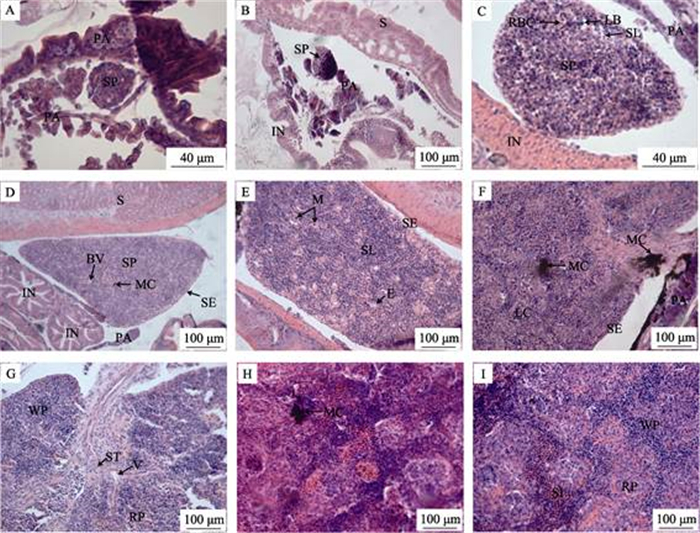
2.3 脾脏7 dph,脾原基出现,位于肠壁背侧,并被胰脏组织包围,卵圆形,由疏松的间充质细胞索和毛细血管组成(图 3A)。16 dph,脾原基增大,梨形,内含少量嗜碱性细胞或造血干细胞,并开始淋巴化(图 3B)。20 dph,脾体积增大,淋巴细胞与红细胞相间排列,可见活跃的红细胞生成;着色较深的嗜碱性细胞、成纤维细胞与红细胞排列较疏松,成索状结构(图 3C)。25 dph,脾外缘由浆膜包裹,淋巴细胞密度增大,毛细血管增多,偶见黑色素巨噬细胞聚集中心(图 3D)。30 dph,脾脏增大,小淋巴细胞增多,巨噬细胞出现,黄褐色,形状不规则;毛细血管增多,椭圆体结构出现(图 3E)。35 dph,黑色素巨噬细胞聚集中心数量增多,体积增大;淋巴细胞和红细胞增多,着色加深,出现明显的淋巴细胞集中区(图 3F)。45 dph,结缔组织被膜伸入脾实质内形成脾小梁,脾小叶开始形成,着色较深的白髄和着色较浅的红髓开始区分,红细胞增多,巨噬细胞明显(图 3G)。53 dph,淋巴细胞数量增加,着色加深,呈紫红色(图 3H),红髓与白髄交错排列,脾窦较明显(图 3I)。此时,脾组织发育基本完成,

|
图 3 四指马鲅脾脏的发育
Fig.3 Development of spleen in E. tetradactylum
A:7 dph,示脾脏原基和胰脏,400×;B:16 dph,脾脏体积增大,细胞开始淋巴化,200×;C:20 dph, 示淋巴母细胞,小淋巴细胞和红细胞,400×;D:25 dph,示黑色素-巨噬细胞聚集中心,浆膜,血管,200×; E:30 dph,小淋巴细胞增多,示巨噬细胞,椭圆体,200×;F:35 dph,示中央静脉,淋巴细胞聚集区,200×; G:45 dph,示脾小梁,红髓,白髄,200×;H:53 dph,200×;I:53 dph,示脾窦,红髓,白髄,200×。 BL:血管;E:椭圆体;IN:肠;LB:淋巴母细胞;LC:淋巴细胞聚集区;M:巨噬细胞; MC:黑色素-巨噬细胞聚集中心;PA:胰脏;RBC:红细胞;RP:红髓;S:胃;SE:浆膜; SP:脾脏;SL:小淋巴细胞;ST:脾小梁;SI:脾窦;V:静脉;WP:白髄 A: 7 dph, showing the spleen primordium and pancreas, 400×; B: 16 dph, showing the spleen bulked up, and the cells began to lymphatize, 200×; C: 20 dph, showing the lymphoblast, small lymphocytes and red blood cells, 400×; D: 25 dph, showing blood vessel, melano-macrophage centers and serosa, 200×; E: 30 dph, showing small lymphocytic increase, macrophage and ellipsoid, 200×; F: 35 dph, showing central veins, lymphocyte aggregation center, 200×; G: 45 dph, showing spleen trabecular, red pulp, white pulp, 200×; H: 53 dph, 200×; I: 53 dph, showing splenic sinus, red pulp, white pulp, 200×. BL: Blood vessel; CV: Central veins; E: Ellipsoid; IN: Intestines; LB: Lymphoblast; LC: Lymphocyte aggregation center; M: Macrophage; MC: Melano-macrophage centers; PA: Pancreas; RBC: Red blood cell; RP: Red pulp; S: Stomach; SE: Serosa; SP: Spleen; SL: Small lymphocytic; ST: Spleen trabecular; SI: Splenic sinus; V: Vein; WP: White pulp |
但尚未成熟,往后继续增大,脾细胞继续淋巴化。脾的内皮系统较头肾发达,其发育速度较胸腺和头肾慢,淋巴细胞明显少于胸腺和头肾。
3 讨论在许多硬骨鱼类发育过程中,胸腺是第一个发育成熟的淋巴器官,如斑马鱼在4 dph时,其胸腺在其他淋巴器官开始发育之前就已经形成(Hansen et al, 1998; Willett et al, 1999)。本研究结果显示,四指马鲅胸腺原基及头肾原基均在3 dph出现,但胸腺原基发育较快,25 dph基本发育完成,而头肾淋巴化速度较慢;脾脏原基则在7 dph形成,发育过程较为缓慢。四指马鲅淋巴器官淋巴化的顺序是胸腺、头肾、脾脏,可见胸腺作为鱼类中枢免疫器官在维系机体早期发育阶段的免疫机能具有重要作用(Rauta et al, 2012)。这与斜带石斑鱼、军曹鱼(Rachycentron canadum)等海水鱼类的研究结果一致(吴金英等, 2003; 苏友禄等, 2008)。四指马鲅的胸腺与鳃咽腔由组织被膜相隔,与咽上皮密切相关,这有利于口咽腔和鳃组织抵抗病原入侵,促进和调节淋巴细胞和非淋巴细胞的相互作用,发挥免疫防御功能(Boehm et al, 2003)。胸腺组织结构的分化在硬骨鱼类中是高度可变的,在许多鱼类中胸腺的皮质和髓质没有明显的分化(Liu et al, 2004)。25 dph胸腺皮质和髓质区分明显,髓质可以看成是一个次级淋巴器官,因为,它可以被外源性抗原和淋巴细胞所利用( Schuurman et al, 1997)。不同的鱼类其免疫能力存在差异可能与胸腺的组织结构特征有一定的关系。
本研究发现,在四指马鲅早期发育过程中,胸腺外周有一些分散的淋巴细胞;胸腺与头肾互相靠拢,有细胞“桥”连接。同样,在金头鲷(Sparus aurata)、罗非鱼(Oreochromis spp)、虹鳉(Poecilia rticulatus)、鲽(Pleuronectes platessa)等鱼类中也有关于胸腺与头肾存在细胞“桥”相连的报道(Jósefsson et al, 1993)。3 dph时,四指马鲅头肾原基中未分化的造血干细胞在形态特征与胸腺的干细胞相似,此时,胸腺和头肾均未开始淋巴化;5 dph时胸腺淋巴细胞有往头肾迁移的迹象,头肾的造血干细胞开始分化,出现淋巴样组织。这为“头肾的淋巴细胞是从胸腺迁移而来”这一观点提供佐证(Jósefsson et al, 1993; Bowden et al, 2005)。但与斜带石斑鱼(吴金英等, 2003)及卵形鲳鲹(Trachinotus ovatus) (蔡文超等, 2012)的研究结果不尽相同,可见不同的鱼类其头肾淋巴细胞的起源存在差别。四指马鲅头肾在发育早期有较多的肾小管,随着鱼体的生长,淋巴细胞增多,18 dph时肾小管开始
退化,直到53 dph肾小管才完全消失,后来主要由淋巴细胞、粒细胞和血细胞等填充,此时头肾形成了具有免疫和造血功能的淋巴样器官。这与牙鲆、斜带石斑鱼、草鱼、鳜(Siniperca chuatsi)、黄颡鱼(Pelteo bagrus fulvidraco)等多种鱼类的研究结果类似(吴金英等, 2003; 雷雪彬等, 2013; 田敬云等, 2005; 刘小玲等, 2009)。20 dph时头肾开始出现少量巨噬细胞和肾上腺细胞,巨噬细胞可吞噬和消灭入侵的病原体,还有助于释放对宿主免疫应答启动起重要作用的促炎细胞因子,表明此时四指马鲅的头肾可能开始具有免疫防御功能(Pressley et al, 2005; Nayak et al, 2007; Fishelson, 2010)。25 dph时头肾淋巴细胞增多,边缘出现大量黄褐色或黑色的嗜铬细胞团,而成熟的嗜铬细胞和肾上腺细胞可分泌儿茶酚胺类激素和皮质类固醇激素,从而参与水盐代谢及应激反应(Geven et al, 2017)。脾脏原基在7 dph时出现,淋巴化的时间最迟,30 dph时有黑色素–巨噬细胞聚集中心出现,往后不断增多,因其有从循环系统中清除可溶性和颗粒物质的作用,常被广泛用作监测水质和鱼体健康状况的生物标记(刘小玲等, 2009)。53 dph时红髓与白髄相间排列,区分不明显,此时,脾脏尚未具备强大的造血和免疫功能,需要继续发育完善。在脾脏发育过程中,红细胞较为活跃,数量明显增长,血管系统发达,表明脾脏具有很强的造血、血液过滤及储存功能(区又君等, 2015)。相对于胸腺和头肾,脾脏淋巴化过程较为缓慢,淋巴细胞也明显少于胸腺和头肾,可见脾脏在体液免疫中处于相对次要的地位(Rauta et al, 2012)。
对于脊椎动物而言,免疫活性的建立依赖于免疫淋巴器官的发育成熟。研究表明,鱼类淋巴细胞的出现并不代表其功能成熟,而淋巴细胞的功能成熟往往比淋巴器官的出现迟,一般要到幼鱼期以后才发育完成,而免疫功能的完善则更迟(吴金英等, 2003)。此外,鱼类淋巴器官的形态及功能发育受营养及环境的影响较大,如环境温度会影响胸腺形态结构的发育以及淋巴细胞的生成(Bowden et al, 2005),矿物质缺乏(如铁、磷、硒)会损害幼鱼免疫器官的功能和结构完整性(Guo et al, 2017; Chen et al, 2017; Lin et al, 2018);拥挤应激会对鱼类的非特异性免疫反应产生负面影响,增加脾脏的不适当凋亡,使鱼类更易受到病原体的侵袭,最终影响鱼类的生存(Zheng et al, 2018)。相对其他鱼类而言,四指马鲅淋巴器官原基出现的时间较早,胸腺淋巴化也较迅速,但头肾和脾脏发育较滞后。四指马鲅从出膜经过仔鱼期、稚鱼期再到幼鱼期需要较长的一段时间过渡。从稚鱼到幼鱼这一变态发育阶段,鱼体的形态结构与器官的发育尚处于演变和完善过程,加上环境因子和营养因素对免疫淋巴器官结构及功能发育的影响,导致其免疫防御机能较弱,这可能是变态期间鱼苗脆弱,容易患病,死亡率高的主要原因。针对该问题亟待进一步往免疫调控机制方面深入研究,这对苗种培育和成鱼健康养殖具有重要意义。
Boehm T, Bleul CC, Schorpp M. Genetic dissection of thymus development in mouse and zebrafish. Immunological Reviews, 2003, 195(1): 15 |
Boehm T, Hess I, Swann JB. Evolution of lymphoid tissues. Trends in Immunology, 2012, 33(6): 315 DOI:10.1016/j.it.2012.02.005 |
Bowden TJ, Cook P, Rombout JHWM. Development and function of the thymus in teleosts. Fish and Shellfish Immunology, 2005, 19(5): 413 DOI:10.1016/j.fsi.2005.02.003 |
Cai WC, Ou YJ, Li JE, et al. Development of immune organs at early stages of Trachinotus ovatus. South China Fisheries Science, 2012, 8(5): 39 |
蔡文超, 区又君, 李加儿, 等. 卵形鲳鲹免疫器官的早期发育. 南方水产科学, 2012, 8(5): 39 DOI:10.3969/j.issn.2095-0780.2012.05.006 |
Campoverde C, Andree KB, Milne DJ, et al. Ontogeny of lymphoid organs and mucosal associated lymphoid tissues in meagre (Argyrosomus regius). Fish and Shellfish Immunology, 2019, 84: 509 DOI:10.1016/j.fsi.2018.09.033 |
Chen K, Jiang WD, Wu P, et al. Effect of dietary phosphorus deficiency on the growth, immune function and structural integrity of head kidney, spleen and skin in young grass carp (Ctenopharyngodon idella). Fish and Shellfish Immunology, 2017, 63: 103 DOI:10.1016/j.fsi.2017.02.007 |
Danilova N, Hohman VS, Sacher F, et al. T cells and the thymus in developing zebrafish. Developmental and Comparative Immunology, 2004, 28(7): 755 |
Espinosa C, García Beltrán JM, Esteban MA, et al. In vitro effects of virgin microplastics on fish head-kidney leucocyte activities. Environmental Pollution, 2018, 235: 30 DOI:10.1016/j.envpol.2017.12.054 |
Fishelson L. Cytomorphological alterations of the thymus, spleen, head-kidney, and liver in cardinal fish (Apogonidae, Teleoste) as bioindicators of stress. Journal of Morphology, 2006, 267(1): 57 DOI:10.1002/jmor.10385 |
Geven EJW, Klaren PHM. The teleost head kidney: Integrating thyroid and immune signalling. Developmental and Comparative Immunology, 2017, 66: 73 DOI:10.1016/j.dci.2016.06.025 |
Guo YL, Jiang WD, Wu P, et al. The decreased growth performance and impaired immune function and structural integrity by dietary iron deficiency or excess are associated with TOR, NF-κB, p38MAPK, Nrf2 and MLCK signaling in head kidney, spleen and skin of grass carp (Ctenopharyngodon idellus). Fish and Shellfish Immunology, 2017, 65: 145 DOI:10.1016/j.fsi.2017.04.009 |
Hansen JD, Zapata AG. Lymphocyte development in fish and amphibians. Immunological Reviews, 1998, 166(1): 199 |
Hu LL, Li JE, Ou YJ, et al. Study on microstructure of head-kedney and speed of Oplegnathus fasciatus. South China Fisheries Science, 2010, 6(3): 41 |
胡玲玲, 李加儿, 区又君, 等. 条石鲷头肾和脾脏的显微结构观察. 南方水产, 2010, 6(3): 41 DOI:10.3969/j.issn.1673-2227.2010.03.008 |
Jósefsson S, Tatner MF. Histogenesis of the lymphoid organs in sea bream (Sparus aurata L. ). Fish and Shellfish Immunology, 1993, 3(1): 35 DOI:10.1006/fsim.1993.1004 |
Lei XB, Chang OQ, Shi CB, et al. Histological and immunohistochemical observations on the early development of head kidney in Ctenopharyngodon idella. Journal of Fisheries of China, 2013, 37(6): 840 DOI:10.3724/SP.J.1231.2013.38530 |
雷雪彬, 常藕琴, 石存斌, 等. 草鱼头肾发生组织学与免疫组织化学观察. 水产学报, 2013, 37(6): 840 |
Lin W, Li L, Chen J, et al. Long-term crowding stress causes compromised nonspecific immunity and increases apoptosis of spleen in grass carp (Ctenopharyngodon idella). Fish and Shellfish Immunology, 2018, 80: 540 DOI:10.1016/j.fsi.2018.06.050 |
Lin XZ, Ou YJ, Li JE, et al. Present status and foreground of family threadfin fishes (Polynemidae). Journal of Biology, 2015, 11061106(4): 83 |
林先智, 区又君, 李加儿, 等. 马鲅科(Polynemidae)鱼类的研究现状及展望. 生物学杂志, 2015, 11061106(4): 83 DOI:10.3969/j.issn.2095-1736.2015.04.083 |
Liu QQ, Wen JF, Ou YJ, et al. The effects of acute off-water handling stress on the tissue structure and oxidative stress of juvenile Eleutheronema tetradactylum. Progress in Fishery Sciences, 2017, 38(6): 48 |
刘奇奇, 温久福, 区又君, 等. 急性离水操作胁迫对四指马鲅(Eleutherone ma tetradactylum)幼鱼组织结构和氧化应激的影响. 渔业科学进展, 2017, 38(6): 48 |
Liu XL, Ge HY, Gu ZM. Ontogeny and structure of head kidney of Pelteobagrus fulvidraco. Journal of Hydroecology, 2009, 2(3): 108 |
刘小玲, 葛海燕, 顾泽茂. 黄颡鱼头肾的组织发生与组织结构研究. 水生态学杂志, 2009, 2(3): 108 |
Liu Y, Zhang SC, Jiang GL, et al. The development of the lymphoid organs of flounder, Paralichthys olivaceus, from hatching to 13 months. Fish and Shellfish Immunology, 2004, 16(5): 621 DOI:10.1016/j.fsi.2003.10.001 |
Ma DC, Ye LH, Xu AY, et al. A histological study of Tylorrhynchus heterochaetus. South China Fisheries Science, 2014, 10(4): 58 |
马定昌, 叶柳荷, 许爱娱, 等. 疣吻沙蚕组织切片的观察研究. 南方水产科学, 2014, 10(4): 58 DOI:10.3969/j.issn.2095-0780.2014.04.010 |
Moore BR, Stapley J, Allsop Q, et al. Stock structure of blue threadfin Eleutheronema tetradactylum across northern Australia, as indicated by parasites. Journal of Fish Biology, 2011, 78(3): 923 DOI:10.1111/j.1095-8649.2011.02917.x |
Nakanishi T, Shibasaki Y, Matsuura Y. T cells in fish. Biology, 2015, 4: 640 DOI:10.3390/biology4040640 |
Nayak AS, Lage CR, Kim CH. Effects of low concentrations of arsenic on the innate immune system of the zebrafish (Danio Rerio). Toxicological Sciences, 2007, 98(1): 118 DOI:10.1093/toxsci/kfm072 |
Ou YJ, Liu JH, Li JE, et al. Morphology and histology of head-kidney and spleen in Cromileptes altivelis. Journal of Southern Agriculture, 2015, 46(11): 2034 |
区又君, 刘江华, 李加儿, 等. 驼背鲈头肾和脾脏的形态组织学观察. 南方农业学报, 2015, 46(11): 2034 DOI:10.3969/j:issn.2095-1191.2015.11.2034 |
Ou YJ, Xie MJ, Li JE, et al. First maturation and mass seedling propagation of cultured Eleutheronema tetradactylum in Guangdong Province. South China Fisheries Science, 2017, 13(4): 97 |
区又君, 谢木娇, 李加儿, 等. 广东池塘培育四指马鲅亲鱼初次性成熟和苗种规模化繁育技术研究. 南方水产科学, 2017, 13(4): 97 DOI:10.3969/j.issn.2095-0780.2017.04.012 |
Pressley ME, PhelanⅢ PE, Witten PE. Pathogenesis and inflammatory response to Edwardsiella tarda infection in the zebrafish. Developmental and Comparative Immunology, 2005, 29(6): 501 DOI:10.1016/j.dci.2004.10.007 |
Rauta PR, Nayak B, Das S. Immune system and immune responses in fish and their role in comparative immunity study: A model for higher organisms. Immunology Letters, 2012, 148(1): 23 |
Schuurman HJ, Kuper CF, Kendall MD. Thymic microenvironment at the light microscopic level. Microscopy Research and Technique, 1997, 38(3): 216 DOI:10.1002/(SICI)1097-0029(19970801)38:3<216::AID-JEMT3>3.0.CO;2-K |
Su YL, Feng J, Guo ZX, et al. Morphological studies on the development of lymphoid organs in Cobia Rachycentron canadum. Marine Fisheries Research, 2008, 29(4): 7 |
苏友禄, 冯娟, 郭志勋, 等. 军曹鱼淋巴器官发育的形态学研究. 海洋水产研究, 2008, 29(4): 7 |
Tian JY, Xie HX, Yao WJ, et al. Ontogenetic development of the pronephros and the distribution of B-lymphocytes in adult pronephros of the mandarin fish Siniperca chuatsi. Acta Zoologica Sinica, 2005, 51(3): 440 |
田敬云, 谢海侠, 姚卫建, 等. 鳜鱼头肾的组织发生及成鱼头肾B淋巴细胞的分布. 动物学报, 2005, 51(3): 440 |
Wang JJ, Peng S, Fei Y. Low mtDNA Cytb diversity and shallow population structure of Eleutheronema tetradactylum in the East China Sea and the South China Sea. Biochemical Systematics and Ecology, 2014, 55(2): 268 |
Willett CE, Cortes A, Zuasti A, et al. Early hematopoiesis and developing lymphoid organs in the zebrafish. Developmental Dynamics, 1999, 214(4): 323 DOI:10.1002/(SICI)1097-0177(199904)214:4<323::AID-AJA5>3.0.CO;2-3 |
Wu JY, Lin HR. Ontogeny of lymphoid organs in the orange- spotted grouper (Epinephelus coioides). Acta Zoologica Sinica, 2003, 49(6): 819 |
吴金英, 林浩然. 斜带石斑鱼淋巴器官个体发育的组织学. 动物学报, 2003, 49(6): 819 |
Xing HF, Gao FT, Zhang YZ, et al. Molecular cloning, expression and SNP screening of natural resistance- associated macrophage protein (Nramp) gene cDNA from half smooth tongue sole (Cynoglossus semilaevis). Progress in Fishery Sciences, 2016, 37(4): 116 |
邢贺飞, 高峰涛, 张永珍, 等. 半滑舌鳎(Cynoglossus semilaevis)基因克隆与表达分析及SNP筛选. 渔业科学进展, 2016, 37(04): 116 |
Zheng L, Jiang WD, Feng L, et al. Selenium deficiency impaired structural integrity of the head kidney, spleen and skin in young grass carp (Ctenopharyngodon idella). Fish and Shellfish Immunology, 2018, 82: 408 DOI:10.1016/j.fsi.2018.08.038 |


